File:PET-image.jpg
From Gomerpedia

Size of this preview: 679 × 600 pixels. Other resolutions: 272 × 240 pixels | 543 × 480 pixels | 869 × 768 pixels | 1,132 × 1,000 pixels.
Original file (1,132 × 1,000 pixels, file size: 139 KB, MIME type: image/jpeg)
Summary
| Description |
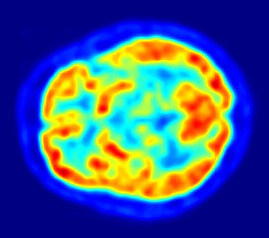
English: This is a transaxial slice of the brain of a 56 year old patient (male) taken with positron emission tomography (PET). The injected dose have been 282 MBq of 18F-FDG and the image was generated from a 20 minutes measurement with an ECAT Exact HR+ PET Scanner. Red areas show more accumulated tracer substance (18F-FDG) and blue areas are regions where low to no tracer have been accumulated.
العربية: صورة مقطعية للدماغ البشري تظهر استهلاك الطاقة. |
||
| Date | |||
| Source | Own work | ||
| Author | Jens Maus (http://jens-maus.de/) | ||
| Permission (Reusing this file) |
|
File history
Click on a date/time to view the file as it appeared at that time.
| Date/Time | Thumbnail | Dimensions | User | Comment | |
|---|---|---|---|---|---|
| current | 19:00, 11 December 2017 | | 1,132 × 1,000 (139 KB) | SteinsplitterBot | Bot: Image rotated by 270° |
File usage
The following 2 pages link to this file: