Difference between revisions of "Bird's Beak"
| Line 8: | Line 8: | ||
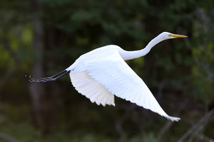
[[File:Egret.jpg|300px|thumb|center|''Picture of whole egret including its beak found on endoscopy January 2014 and released shortly thereafter back into the wild'']] | [[File:Egret.jpg|300px|thumb|center|''Picture of whole egret including its beak found on endoscopy January 2014 and released shortly thereafter back into the wild'']] | ||
| + | |||
| + | [[Category: Gastroenterology]] | ||
Revision as of 09:53, 16 July 2016
The bird's beak refers to not only one but two findings in patients with achalasia. Classically, a bird's beak appearance is noted along with a dilated esophagus on barium swallow. The lesser known but truer bird's beak is when upper endoscopy reveals an actual bird's beak and sometimes a whole bird's head lodged at the GE (gastroesophageal) junction thus causing dysphagia and regurgitation.
Treatment
The treatment for a true bird's beak on endoscopy is to remove the bird's beak and other bird parts. In rare instances, you might find a whole live bird. Do not forget to send to pathology to determine what type of bird it is. Pigeons are the most common, though rare bird's beaks include the Asian crested ibis, red-crowned crane, and orange-bellied parrot.

